Minimally Invasive Aortic Valve Surgery
 "UCSF offers the full range of minimally invasive approaches for aortic valve repair and replacement, a
 What is Aortic Valve Disease?
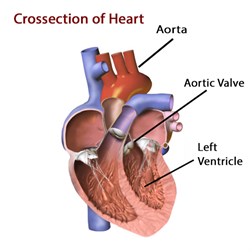
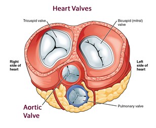
The aortic valve consists of three leaflets or cusps and is located between the left ventricle of the heart and the aorta. The aortic valve prevents blood from leaking back into the heart after it has been ejected during the contraction. When the valve fails, patients may be severely affected. The aortic valve is one of the four major valves in the heart in addition to the tricuspid or right atrioventricular valve, mitral valve, and pulmonary value.
 |
 |
| By OpenStax College [CC BY 3.0 ], via Wikimedia Commons |
"Medical gallery of Blausen Medical 2014". WikiJournal of Medicine 1 (2). |
Aortic Valve Stenosis
The term stenosis means a narrowing. In aortic valve stenosis, the aortic valve becomes narrowed, stiff or thickened impairing the ejection of the blood which is necessary to the proper functioning of the heart. Aortic valve stenosis is the most common valvular heart defect in the U.S. affects nearly 2% of people over 65 years of age or nearly 1 million people.
Symptoms
- Decreased ability to exercise
- Shortness of breath
- Loss of consciousness or chest pain In advanced disease
Causes
There are multiple sources of aortic valve stenosis
- Bicuspid Valve Malformation - this is an inherited defect in which two of the leaflets of the aortic valve fuse during development resulting in a two-leaflet valve called bicuspid valve malformation, vs. the normal three-leaflet (tricuspid) valve
- Rheumatic Valvular Disease - the aortic valve leaflets fuse and become fibrotic. This condition is most commonly seen in the developing world.
-
Calcified Aortic Valve Disease - the three-leaflet valve becomes calcified through a buildup of calcium deposits which can harden effectively narrowing the valve. This is most common cause of aortic stenosis in adults and is thought to be a degenerative process.
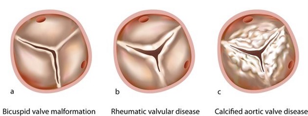 |
Aortic Value Disease - a) In bicuspid valve malformation, two of the leaflets of the aortic valve fuse during development resulting in a two-leaflet valve instead of the normal three-leaflet (tricuspid) valve b) Here the aortic valve leaflets fuse and become fibrotic causing rheumatic heart disease c) The most common cause of aortic stenosis in adults is calcification of the normal three-leaflet valve, likely a degenerative process |
Aortic Valve Insufficiency
In aortic valve insufficiency, the aortic valve fails to close properly causing the ejected blood to leak back into the heart.
Symptoms
Symptoms of aortic insufficiency are similar to those of heart failure and may include:
- Decreased ability to exercise
- Shortness of breath
- Heart palpitations
- Chest pain
Why is an Aortic Valve Defect Dangerous?
Patients may become symptomatic, experiencing shortness of breath, reduced exercise capacity, fatigue, or may even become unconscious and fall. Severe aortic valve defects may result in heart failure and reduces life expectancy.
When Should Aortic Valve Defects be Treated?
Studies have shown that severe aortic valve defects (stenosis or insufficiency) should be treated before irreversible structural changes in the heart occur. In certain forms of aortic valve insufficiency, the aortic valve can be repaired; most of the stenotic aortic valves need replacement.
Minimally-Invasive Aortic Value Surgery
Traditionally, aortic valve surgery is performed through a large mid-line skin incision in which the breastbone is divided called a sternotomy. However, in patients with isolated aortic valve disease who do not have symptoms of widespread coronary artery disease, the surgery may be performed using minimally invasive techniques.
Surgical Procedure
The surgeon makes a 2-3 inch incision on the right side of the chest, a procedure called a mini-thoracotomy, or a similarly sized smaller incision using a hemi-sternotomy, a lesser operation in which the incision is used to divide only the upper half of the breastbone.
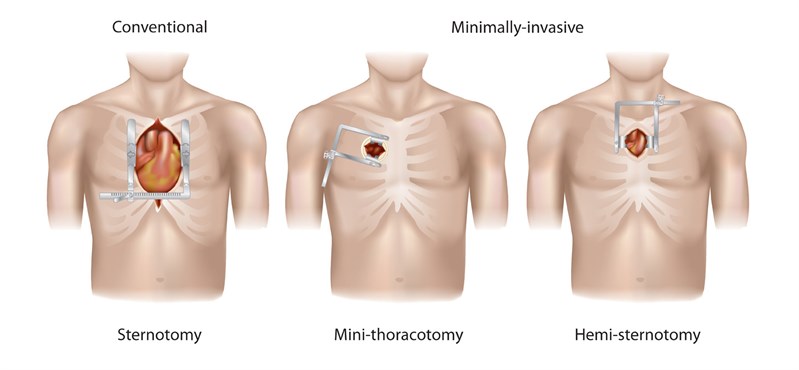 |
Surgical Approaches - In traditional aortic valve surgery, the surgeon gains uses a sternotomy to gain access to the aortic value requiring the spreading of the breastbone. Where minimally invasive techniques are used, either via a mini-thoracotomy or hemi-sternotomy, a much lesser operation in terms entry to the operative field can be performed. |
A heart-lung machine is then inserted either via a small incision in the groin or as described above. This allows the heart to be stopped during the replacement of the valve with the heart-lung machine taking over the normal functions of the heart.
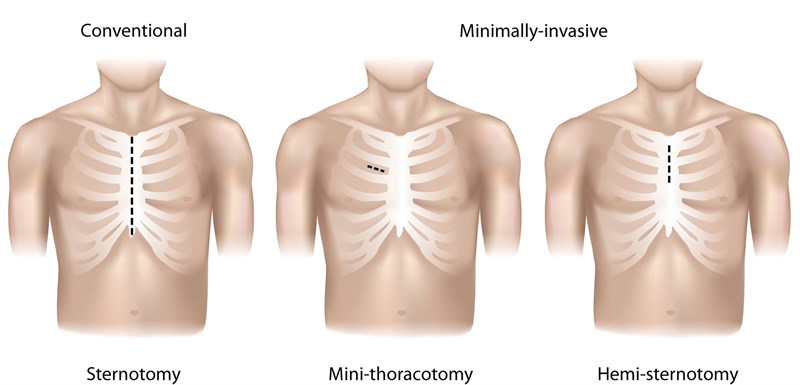 |
Types of Incisions - In traditional aortic valve surgery, the surgeon makes a large 4-6 inch incision to gain access to the aortic value and spreads the breastbone. With either a mini-thoracotomy or hemi-sternotomy, a much lesser operation is required using only a 2-3 inch incision. |
Benefits of Minimally Invasive Aortic Valve Surgery
There are considerable advantages to performing aortic value surgery with minimally invasive techniques including:
- A smaller scar and improved appearance
- Reduced blood loss and risk of infection during and after the surgery
- Reduced post-surgical pain and reliance on pain medications
- Faster recovery from surgery
- Shorter hospital stay
- Shorter post-operative recovery with a quicker return to normal activities


